


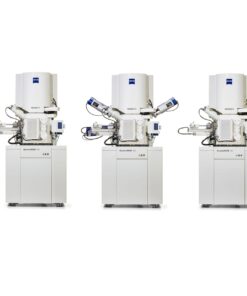
Revolutionize the Speed of Electron Microscopy
Multiple electron beams working in parallel give you unprecedented gross imaging speed. Acquiring an area of 1 mm2 at 4 nm pixel size takes only a few minutes. The unrivaled acquisition speed of more than 1 TB per hour enables imaging of large volumes (> 1 mm3) at nanometer resolution. Optimized detectors collect the secondary electron signals very efficiently, providing you with high contrast images at low noise levels.
Image Huge Samples at Nanometer Resolution
Don’t sacrifice sample size for nanometer resolution. MultiSEM is equipped with a sample holder covering an area of 10 cm × 10 cm and built for continuous 24/7 operation. You can finally image the entire sample and discover everything you need to answer your scientific questions. You get the detailed picture‚ without losing the macroscopic context.
Electron Microscopy with ZEN Imaging Software
By introducing ZEN to MultiSEM, we bring the standard software for ZEISS light microscopes to the world of electron microscopy. Control MultiSEM in a straightforward, intuitive way: Smart auto-tuning routines support you as you capture optimal images with high resolution and quality. You quickly set up even complex automated acquisition procedures, adapted and tuned to your sample imaging.


 VI
VI