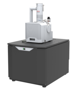
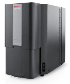
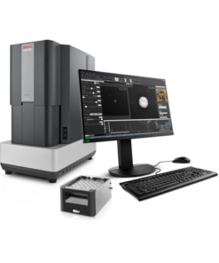
Volumescope 2 Scanning Electron Microscope for serial block face imaging
The Thermo Scientific Volumescope 2 Scanning Electron Microscope (SEM) is our state-of-the-art serial block-face imaging system. Keep control of experiments with easy to use technology and protect a wide range of possible samples with tried and tested solutions for every acquisition step.
Block face imaging
Unraveling the complex 3D architecture of cells and tissues in their natural context is crucial for structure-function correlation in biological systems and soft materials. Serial block-face SEM (SBF-SEM) combines in situ sectioning and imaging of plastic-embedded tissue blocks within the SEM vacuum chamber for automated, 3D reconstruction of large tissue volumes. Axial resolution was once limited by the minimum section thickness, but truly isotropic resolution is now possible with the addition of multi-energy deconvolution.
3D SEM
Following in situ sectioning of the block-face using a diamond knife, the freshly exposed tissue is imaged several times using increasing accelerating voltages. These images are subsequently used in a deconvolution algorithm to derive several optical subsurface layers, forming a 3D subset. By repeating this cycle, the Volumescope 2 SEM offers isotropic datasets with 10 nm Z-resolution.
Polymer microstructures
High-resolution imaging in 3D is of great importance when trying to understand the microstructure and properties of polymer materials. The Volumescope 2 SEM combines in situ sectioning and imaging of polymers within the SEM vacuum chamber, in a fully automated fashion, for reconstruction of large volumes with truly isotropic 3D resolution. Visualizing a large volume at such resolutions is critical for revealing how regions with different microstructures affect the overall properties of the material. For example, the Volumescope 2 SEM can recreate a large representative volume of a filtration membrane, providing accurate transport properties that lead to enhanced performance predictions for new filter designs.
Easy to use technology
Keep control of experiments with easy to use technology. Reuse jobs and system settings and select multiple regions of interest during job acquisition.
Visualize and navigate during acquisition
Visualize and navigate during acquisition with Thermo Scientific Amira Live Tracker Software to optimize/control your outcome/Automate large 3D volume acquisitions as well as reconstructions.
Protect valuable samples
Protect valuable samples with tested solutions at every acquisition step: debris trap and swipe features ensure sample quality; low vacuum detector enables imaging of highly charged samples.
Easy and fast mounting microtome exchange
Easy and fast mounting microtome exchange for normal SEM operation or automated array tomography with the addition of Thermo Scientific Maps Software.
Pathology Research
Transmission electron microscopy (TEM) is used when the nature of the disease cannot be established via alternative methods. With nano-biological imaging, TEM provides accurate and reliable insight for certain pathologies.
Plant Biology Research
Fundamental plant biology research is enabled by cryo electron microscopy, which provides information on proteins (with single particle analysis), to their cellular context (with tomography), all the way up to the overall structure of the plant (large volume analysis).
Fundamental Materials Research
Novel materials are investigated at increasingly smaller scales for maximum control of their physical and chemical properties. Electron microscopy provides researchers with key insight into a wide variety of material characteristics at the micro- to nano-scale.
Quality control and failure analysis
Quality control and assurance are essential in modern industry. Thermo Fisher offers a range of EM and spectroscopy tools for multi-scale and multi-modal analysis of defects, allowing you to make reliable and informed decisions for process control and improvement.
A novel serial block-face imaging (SBFI) solution that combines multi-energy deconvolution scanning electron microscopy (MED-SEM) with in situ sectioning. Automation and ease-of-use functions provide isotropic resolution for large volume samples.


 VI
VI