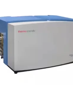
Talos L120C G2 Transmission Electron Microscope
The Thermo Scientific Talos L120C G2 (S)TEM is a 20-120 kV thermionic (scanning) transmission electron microscope uniquely designed for performance and productivity. It’s useful across a wide range of samples and applications, such as 2D and 3D imaging of cells, cell organelles, asbestos, polymers, and soft materials, both at ambient and cryogenic temperatures. The Talos L120C allows you to acquire high-quality results with minimal effort, no matter your skill level. Routine 2D imaging of samples is facilitated by a simple and intuitive user interface (UI). By implementing fast and sophisticated automation in advanced 3D imaging workflows with superior TEM and STEM resolution for 20-120 kV instrumentation, it allows you to focus on scientific questions rather than microscope operation.
Talos L120C (S)TEM) advantages
The Talos L120C takes imaging to the next level with the optional cryo-box and low-dose techniques, which facilitate imaging of samples preserved at cryogenic temperatures and produce high-quality images even for beam-sensitive materials. The cryo-box provides maximal protection of the cryo-specimen resulting in minimal ice growth contamination over long (>8 h) data collection sessions.
To enhance productivity and application flexibility, especially in multi-user and multi-application environments, the cryo-box is motorized and fully retractable to facilitate seamless switching between cryo-EM imaging, STEM imaging, and EDS analysis. The large pole-piece gap of the C-Twin lens provides high contrast and allows for high specimen tilts up to 90°, while the constant-power objective lens with low-hysteresis allows for straightforward and reproducible changes of imaging modes and high-tension settings in matter of seconds.
Overall, the Talos L120C (S)TEM combines application versatility with a reproducibly performing electron column to give you new opportunities for high-resolution 3D characterization, in situ dynamic observation with EDS and other diffraction applications, and a special emphasis on high-contrast imaging and cryo-TEM.\
Talos L120C (S)TEM automation
Designed for multi-user and multi-discipline environments and equipped with a familiar UI shared across all Thermo Scientific TEM platforms, the Talos L120C TEM is ideal for novice and expert users alike. All daily TEM tunings have been automated to provide the best and most reproducible setup, easing the learning curve for novice operators and improving time-to-data for experienced users. The Talos L120C TEM also offers educational online help embedded within the UI. Additionally, Thermo Scientific application software packages facilitate intuitive settings and automated data collection for different use cases and workflows, such as large area 2D imaging, energy dispersive X-ray analysis, single particle analysis, MicroED and tomography.
| TEM line resolution |
|
| TEM point resolution |
|
| STEM HAADF resolution |
|
| TEM magnification range |
- Standard: 25–650k ×
- Enhanced: 35–910k ×
|
| STEM magnification range |
|
| Maximum tilt angle (stage) |
|
Versatile multi-application system
Designed for routine TEM and STEM imaging and characterization of a variety of samples, including resin embedded cells, soft materials, nanoparticles, and protein complexes at ambient and cryo-conditions.
High quality images
High-contrast, high-quality TEM and STEM imaging by simultaneous, multiple signal detection with up to four-channel-integration STEM detectors.
Cryo-imaging
Retractable, motorized cryo-box and low-dose technique enable superior imaging of samples at cryogenic temperatures while preserving the possibility of STEM imaging and EDS analysis applications.
Chemical composition data
Flexible energy-dispersive X-ray spectroscopy (EDS) analysis for chemical information.
Space for more
Add tomography or in situ sample holders compatible with a large analytical pole piece gap allowing up to ±90° stage tilt range and ±375 µm stage height range.
Improved productivity and reproducibility
Ultra-stable column, SmartCam remote operation, and constant power objective lenses for quick mode and high-tension switches. Fast, easy switching for multi-user environments.
Auto-alignments
All daily TEM tunings—such as focus, Eucentric height, center beam shift, center condenser aperture, and rotation center—are automated.
Automated data collection
Automate single particle analysis imaging with Thermo Scientific EPU Software and tomography with Thermo Scientific Tomo 5 Software. Collect large area 2D images at different image resolutions using Thermo Scientific Maps Software with automatic image acquisition and stitching while documenting the overall context and entire area of the region of interest with exceptional quality.
Ceta CMOS Camera
The 4K × 4K Thermo Scientific Ceta CMOS Camera with large field of view enables live digital zooming with high sensitivity and high speed over the entire high-tension range. Specialized Ceta Cameras are available for low-dose imaging (Ceta-S and Ceta-F) and for MicroED (Ceta-D) applications.
Compact design
Smaller footprint and dimensions mean this tool can be accommodated into more challenging spaces while reducing infrastructure and support costs.
Infectious Disease Research
Cryo-EM techniques enable multiscale observations of 3D biological structures in their near-native states, informing faster, more efficient development of therapeutics.
Structural Biology Research
Cryo-electron microscopy enables the structural analysis of challenging biological targets such as large complexes, flexible species and membrane protein.
Drug Discovery
Take advantage of rational drug design for many major drug target classes, leading to best-in-class drugs.
Plant Biology Research
Fundamental plant biology research is enabled by cryo electron microscopy, which provides information on proteins (with single particle analysis), to their cellular context
Pathology Research
Transmission electron microscopy (TEM) is used when the nature of the disease cannot be established via alternative methods.
Quality control and failure analysis
Quality control and assurance are essential in modern industry. We offer a range of EM and spectroscopy tools for multi-scale and multi-modal analysis of defects, allowing you to make reliable and informed decisions for process control and improvement.
Fundamental Materials Research
Novel materials are investigated at increasingly smaller scales for maximum control of their physical and chemical properties. Electron microscopy provides researchers with key insight into a wide variety of material characteristics at the micro- to nano-scale
Process control using electron microscopy
Modern industry demands high throughput with superior quality, a balance that is maintained through robust process control. SEM and TEM tools with dedicated automation software provide rapid, multi-scale information for process monitoring and improvement.
Energy Dispersive Spectroscopy
Energy dispersive spectroscopy (EDS) collects detailed elemental information along with electron microscopy images, providing critical compositional context for EM observations. With EDS, chemical composition can be determined from quick, holistic surface scans down to individual atoms.
3D EDS Tomography
Modern materials research is increasingly reliant on nanoscale analysis in three dimensions. 3D characterization, including compositional data for full chemical and structural context, is possible with 3D EM and energy dispersive X-ray spectroscopy.
EDS Elemental Analysis
Thermo Scientific Phenom Elemental Mapping Software provides fast and reliable information on the distribution of chemical elements within a sample.
Atomic-Scale EDS
Atomic-resolution EDS provides unparalleled chemical context for materials analysis by differentiating the elemental identity of individual atoms. When combined with high-resolution TEM, it is possible to observe the precise organization of atoms in a sample.
Electron Energy Loss Spectroscopy (EELS)
Materials science research benefits from high-resolution EELS for a wide range of analytical applications. This includes high-throughput, high signal-to-noise-ratio elemental mapping, as well as probing of oxidation states and surface phonons.
In Situ experimentation
Direct, real-time observation of microstructural changes with electron microscopy is necessary to understand the underlying principles of dynamic processes such as recrystallization, grain growth, and phase transformation during heating, cooling, and wetting.
Particle analysis
Particle analysis plays a vital role in nanomaterials research and quality control. The nanometer-scale resolution and superior imaging of electron microscopy can be combined with specialized software for rapid characterization of powders and particles.
Automated Particle Workflow
The Automated NanoParticle Workflow (APW) is a transmission electron microscope workflow for nanoparticle analysis, offering large area, high resolution imaging and data acquisition at the nanoscale, with on-the-fly processing.


")


 VI
VI