

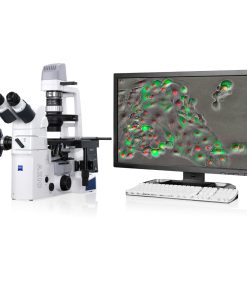
With Primovert HDcam and the imaging App Labscope you observe and discuss cells independently from the sterile workplace together with your colleagues.


Place ZEISS Primovert right inside your Laminar Flow Box. Examine unstained cells in phase contrast and GFP-labeled cells in fluorescence contrast quickly and efficiently.
The inverted microscope is especially perfect for cancer and genetic research.
With Primovert HDcam and the imaging App Labscope you observe and discuss cells independently from the sterile workplace together with your colleagues.
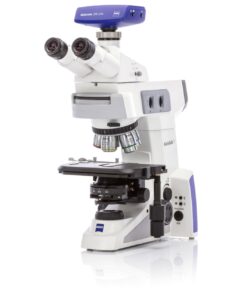
| Objective change | Manual via quadruple objective nosepiece |
| Objectives | Infinity-corrected objective range with W 0.8 mounting thread |
| Plan-Achromat: 4x/0.1, 4x/0.1 Ph0, 10x/0.25 Ph1 | |
| LD Plan-Achromat: 20x/0.3 Ph1, 40x/0.5 Ph1, 20x/0.3 Ph2, 40x/0.5 Ph 2 | |
| Phase-Slider | Universal phase slider for the objectives Ph1: Convenient and economical |
| Phase slider for Ph2: Higher resolution | |
| Eyepieces | WF-PL 10x/20 Br. foc. |
| Specimen stage | Fixed |
| Dimensions (width x depth) | 200 x 239 mm |
| Specimen guide | Right side |
| Verniers with numerical and alphabetic scale Coaxial drive | X direction: Numerical scale, readable from right to left |
| Y direction: Alphabetic scale, readable in the mirror right side | |
| LD condenser 0.3 | For magnifications 4 x to 40 x, WD = 72 mm |
| LD condenser 0.4 | For magnifications 4 x to 40 x, WD = 55 mm |
| Binocular tube 45°/20 Interpupillary distance Viewing height | Viewing angle 45°, FOV 20 Adjustable from 48 to 75 mm 360 to 397 mm |
| Trinocular (photo)tube 45°/20 | Viewing angle 45°, FOV 20 |
| Photo/video port | Tube factor 1x, 60 mm |
| Fixed beam splitting | 50% vis / 50% doc |
| Primo Vert Monitor | Camera: 5 Megapixel CMOS |
| Monitor size: 8.4” | |
| Display: 800 x 600 pixel | |
| Storage Medium: Secure Digital (SD) card | |
| Outputs/ports: USB 2.0 | |
| Camera driver for Microscopy software: AxioVision LE with special Configuration tool | |
| Light source | HAL: 6V,30W |
| LED: White light, 3 W |
Inverted Microscopes
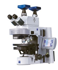
Upright microscopes
Upright microscopes
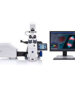
Confocal Microscopes
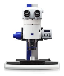
Stereo Microscopes
Upright microscopes
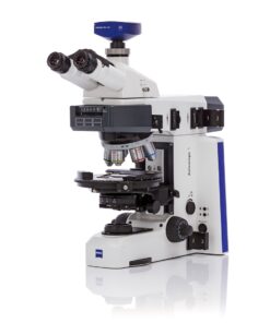
Axioscope 5 – Smart Microscope for Research and Routine in the Materials Lab
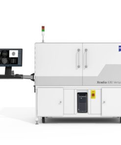
X-ray Microscopes
Camera

 VI
VI