Online Support

Aperio GT 450 – Automated, High Capacity Digital Pathology Slide Scanner

Fast-Forward to Next Generation Imaging

The Aperio GT 450 enables histotechnicians to complete scanning tasks quickly and with confidence leveraging a 32-second scan speed*. Output 81 slides/hr at 40x* delivering high quality images with Leica optics and with an IT architecture that is secure and scalable. From the pathology lab to the IT room, the Aperio GT 450 is designed to scale up digital pathology operations.

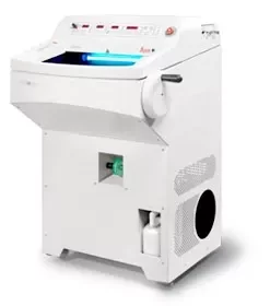
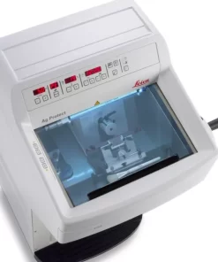
Fast and Easy to Operate for Histotechnicians

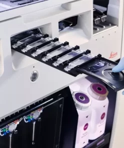
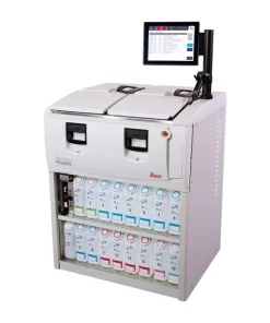
The Aperio GT 450 is simple to operate and helps histotechnicians deliver rapid results with confidence. Complete scanning quickly and easily so you can focus on other tasks in the lab: 15 racks of 30 slides (450 slides total) can be loaded directly from HistoCore SPECTRA CV Coverslipper into the Aperio GT 450. The slides are then automatically scanned at 81 slides per hour at 40x for a 15mm x 15mm area.

  • No touch continuous loading during scanning
  • Automated IQ checks during each scan to ensure quality
  • Assign priority cases
  • Every slide is calibrated with each scan
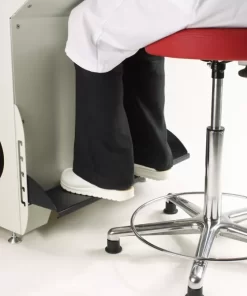
  • 450 slide capacity packed into a small size fits on the lab bench
  • 99.5% accurate tissue finder that allows you to adjust if needed*
  • 100% first-time barcode scan success rate*

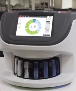
Secure, Scalable Architecture for IT Professionals

The Aperio GT 450 offers fast, secure, and flexible IT architecture. A centralized Scanner Admin Manager (SAM) server and dedicated software provides you with a data management solution that can remotely set up and monitor multiple Aperio GT 450s at a time.*

  • Comprehensive cyber security including encryption and login controls
  • Centralized scanner management to set up and monitor multiple scanners
  • Two 10GB network connections on the SAM server
  • Scalable set up for hub and spoke configurations
  • HL7 compatible for easy integration into LIS, LIMS or PACS
  • Supports dedicated logins for each Aperio GT 450 scanner

Excellent Image Quality for Pathologists

The Aperio GT 450 uses a high-performance objective manufactured by Leica Microsystems, that has produced world class optics since 1847. This historic craftmanship is now built into the today’s Aperio GT 450, which is designed to rapidly deliver images with exceptional quality for Pathologists.

  • Leica optics deliver exceptional image quality
  • Every slide is calibrated during scanning to ensure the best quality image
  • Automated image quality assessment so you don’t have to send slides back for re-scans
  • 99.5% accurate tissue finder finds faint tissue while excluding pen marks, dust, and residue
IMAGING, CAPACITY & OPERATIONS
Magnification:
40x
Scan Speed: 32 sec/slide (@ 40x for 15mm x 15mm area)
Scan Throughput: 81 slides/hr (@40x for 15mm x 15mm area)*
Scan Resolution: 0.26 µm/pixel at 40x
Scan Output: DICOM compatible and SVS
Scanning Region: ≤ 23.6 mm x 58 mm
Field of View (FOV): 1mm
Objective Lens: Custom-made by Leica Microsystems
*VP-0450
Slide Capacity:
450 slides (15 racks of 30)
Slide Loading: Automatic Continuous Load up to 450 1-inch x 3-in (2.54 cm x 7.62 cm) slides
Racks Accepted: Leica Universal Rack (30-slide capacity), validated and recommended for use with HistoCore SPECTRA Workstation
Sakura Prisma Stainer and Coverslipper Rack (20-slide capacity)
DIMENSIONS & WEIGHT
Dimensions (W x L x H):
52.83 cm x 58.43 cm x 49.53 cm
(20.8 in x 23 in x 19.5 in)
Weight:
140 lbs (63.5 kg)
IT REQUIREMENTS & INTEGRATION
Network Bandwidth Requirements (Aperio GT 450 to SAM Server):
1 gigabits per second (Gbps)
Network Bandwidth Requirements (SAM Server to image repository/DSR): 10 gigabits per second (Gbps)
Compatibility:
HL7 and DICOM (for LIS, LIMS, PACS integration)
Image File Formats: DICOM compatible images, SVS, TIFF
SECURITY FEATURES
SSL for SAM User Interface
Assign User Specific PINs per Scanner  –includes automatic timeouts
Secure Sign-On to SAM Server Software
Log Data & Events from Mirth Connect to File System
Remote Access to Scanner Status
Create User Groups and Assign Permissions  –password for client software and PIN for scanner
SLIDES, COVERSLIPS & LABELS
SLIDES
General: The tissue/smear is embedded between supported slide and coverslip
Standard: 1-inch x 3-inch (2.54 cm x 7.62 cm) microscope slides. Measurements comply with ISO 8037/1.
Minimum Slide Size (W x L): 25 mm x 75 mm
Maximum Slide Size (W x L): 26 mm x 76 mm
Thickness: Optimized for range of 0.9 mm to 1.1 mm, excluding coverslip
Special Instructions: The coverslip/label shall not protrude beyond the edge of the glass microscope slide. The entire coverslip and label must be glued to the glass microscope slide. There must be no lifted edges or parts of the coverslip/label. The outer surface of the slide must be dry. No liquid shall flow out of the specimen area.
Slide Preparation: Slides are prepared typically using:
Glass coverslip with mounting media, Eukitt or water
Film coverslip with integrated glue
Maximum Tissue/Smear Thickness: Optimized for tissue thickness of 3-5 μm (including mounting media)
COVERSLIPS
Coverslips Accepted: Optimized for coverslip with thickness of .17 mm, made of typical coverslip material: Standard microscope cover glass or Cellulose Tri-Acetate film (microscope cover film).
LABELS
Label Area (W x L): 25 mm x 25 mm. Handwritten/printed non-transparent, matt (paper-like reflecting) sticker.
Label Special Instructions: Labels shall not protrude beyond the edge of the slides or be lifted. Labels shall not be attached to the bottom of the slide, but only attached to the coverslip-side of the slide.
BARCODES
Barcodes Engines: 1D and 2D Symbology
Barcodes Supported: NW7, QR Code, Data Matrix, Interleaved 2 of 5, Code 39, Code 128, PDF417, MicroPDF417