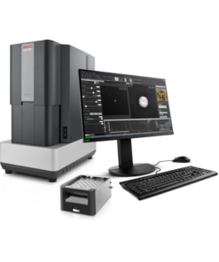
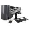
Phenom Pure Desktop SEM
The Thermo Scientific Phenom Pure Desktop Scanning Electron Microscope (SEM) is an ideal tool for the transition from light optical to electron microscopy. It is an economical solution for high-resolution imaging, providing the best imaging results in its class.
The Phenom Pure Desktop SEM provides high-quality images thanks to the long-lasting, high-brightness CeB6 source, and offers the market’s fastest loading and imaging time. The very reliable autofocus and automated source alignments make this the most user-friendly system on the market.
Never lost navigation
The user always knows the position on the sample with the unique never lost navigation. Overviews of both the optical and electron optical images provide clear reference points at all times. Thanks to the integrated motorized stage, you can navigate quickly through your sample.
Easy to use
Users are ready to take images after only 10 minutes of basic training. A large variety of sample holders is available to accommodate a large range of samples. Sample loading is fast and safe due to our patented sample vacuum loading technology.
Customize your SEM
The Phenom Pure Desktop SEM can be equipped with two optional detector systems. The first one is a fully integrated energy dispersive spectroscopy (EDS) system. The second is a secondary electron detector (SED) for applications that require surface and topography sensitive imaging.
Process control using electron microscopy
Modern industry demands high throughput with superior quality, a balance that is maintained through robust process control. SEM and TEM tools with dedicated automation software provide rapid, multi-scale information for process monitoring and improvement.
Quality control and failure analysis
Quality control and assurance are essential in modern industry. We offer a range of EM and spectroscopy tools for multi-scale and multi-modal analysis of defects, allowing you to make reliable and informed decisions for process control and improvement.
Fundamental Materials Research
Novel materials are investigated at increasingly smaller scales for maximum control of their physical and chemical properties. Electron microscopy provides researchers with key insight into a wide variety of material characteristics at the micro- to nano-scale.
EDS Elemental Analysis
Thermo Scientific Phenom Elemental Mapping Software provides fast and reliable information on the distribution of chemical elements within a sample.
3D EDS Tomography
Modern materials research is increasingly reliant on nanoscale analysis in three dimensions. 3D characterization, including compositional data for full chemical and structural context, is possible with 3D EM and energy dispersive X-ray spectroscopy.
Atomic-Scale Elemental Mapping with EDS
Atomic-resolution EDS provides unparalleled chemical context for materials analysis by differentiating the elemental identity of individual atoms. When combined with high-resolution TEM, it is possible to observe the precise organization of atoms in a sample.
Imaging Hot Samples
Studying materials in real-world conditions often involves working at high temperatures. The behavior of materials as they recrystallize, melt, deform, or react in the presence of heat can be studied in situ with scanning electron microscopy or DualBeam tools.
In Situ experimentation
Direct, real-time observation of microstructural changes with electron microscopy is necessary to understand the underlying principles of dynamic processes such as recrystallization, grain growth, and phase transformation during heating, cooling, and wetting.


 VI
VI