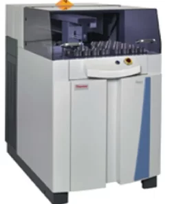
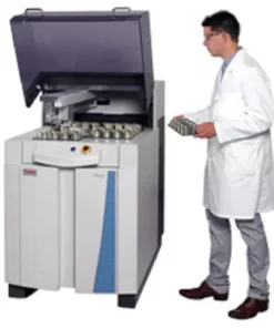
The Thermo Scientific Aquilos 2 Cryo-FIB is the latest generation of our cryo-DualBeam system. It is dedicated to the preparation of thin, electron-transparent lamellas for high-resolution cryo-electron tomography or MicroED of micro-crystals.
With the Aquilos 2 Cryo-FIB, you can take a revolutionary step forward by automating steps in the workflow to produce multiple lamellas from specific programmed locations. The improved dedicated cryo-hardware will provide you with longer run times at ultra-low contamination rates. Discover how our latest cryo-FIB platform enables the determination of structures right inside the cell.
The tomography of biomolecules in their cellular context requires highly accurate sample preparation with cryo-FIB, which can be achieved with information from integrated fluorescence microscopy. Now available for the Aquilos 2 Cryo-FIB, the Thermo Scientific iFLM Correlative System combines light and electron microscopy in one system. This eliminates the need for extra sample transfer steps and enables you to create a more streamlined cryo-correlative solution for cryo-tomography. The iFLM (Integrated Fluorescence Light Microscope) Correlative System localizes fluorescent targets inside the chamber, allowing you to correlate two imaging modalities within one system, thereby ensuring the sample is in the right location.
Automated batch cryo-lamella milling
Mark multiple points of interest, such as cells, for cryo-lamella milling. Cryo AutoTEM Software is a guided solution for automatically preparing multiple lamellas in unattended runs, increasing your productivity.
Cryo-lift-out lamella preparation
Prepare lamellas from specific targeted regions with nanometer-position accuracy. The high-precision EasyLift NanoManipulator option allows for extraction of site-specific regions, such as fluorescently labeled proteins, even from high-pressure frozen samples. These lamellas can subsequently be placed inside autogrids for cryo-TEM and further imaging of protein structures and protein networks.
3D visualization
With our Cryo-Auto Slice and View Software, you can acquire 3D images from vitrified samples by sequential milling and then imaging of the cross-section of a defined volume of the sample. This process will help you stop milling at the correct subcellular region to prepare your lamella before you observe the ultrastructural detail in your cryo-TEM using cryo-ET.
Correlate light and electron within one system
The iFLM Correlative System allows samples to be imaged directly within the high vacuum of the Aquilos 2 Cryo-FIB without additional transfer steps from an external cryo-light microscope. This saves time and reduces the risk of sample contamination. Fluorescently marked cells can be located directly in the Aquilos.
Accelerate and Advance service packages for Cryo-tomography
Accelerate and Advance service packages for Cryo-tomography will support your success from the moment your purchase your Thermo Scientific Aquilos 2 Cryo-FIB system. From applications support to system remote monitoring to comprehensive maintenance, our unique blend of data-driven and hand-on services will help you quickly achieve your desired outcomes.
Preparation of cells for cryo-electron tomography
The Thermo Fisher cryo-electron tomography workflow covers flash freezing cells to final 3D visualization. Interior cellular regions are imaged at nanometer-scale resolution and from cryo-lamellae precisely prepared with Aquilos 2 cryo-FIB.
Infectious Disease Research
Cryo-EM techniques enable multiscale observations of 3D biological structures in their near-native states, informing faster, more efficient development of therapeutics.
Structural Biology Research
Cryo-electron microscopy enables the structural analysis of challenging biological targets such as large complexes, flexible species and membrane protein.
Drug Discovery
Learn how to take advantage of rational drug design for many major drug target classes, leading to best-in-class drugs.
Cryo-Tomography
Cryo-electron tomography (cryo-ET) delivers both structural information about individual proteins as well as their spatial arrangements within the cell. This makes it a truly unique technique and also explains why the method has such an enormous potential for cell biology. Cryo-ET can bridge the gap between light microscopy and near-atomic-resolution techniques like single-particle analysis.
")


 VI
VI