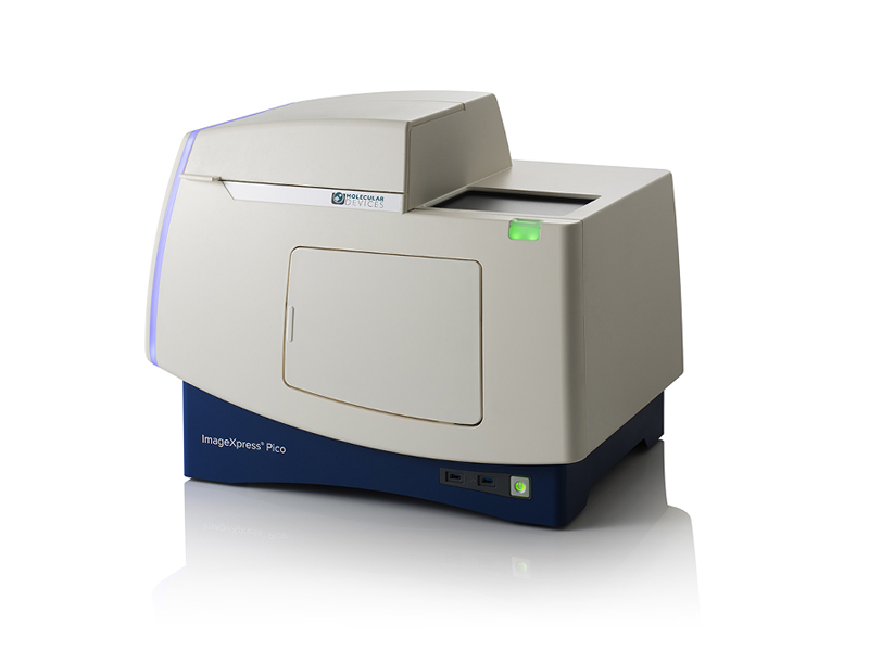
Digital microscopy with automated brightfield, fluorescence, and Digital Confocal imaging
The ImageXpress® Pico Automated Cell Imaging System is more than a digital microscope, combining high-resolution imaging with powerful analysis. Whether running fluorescence imaging or brightfield assays, the automated imager features a comprehensive portfolio of preconfigured protocols for cell-based assays to shorten the learning curve, so you can start running experiments quickly. With features such as Digital Confocal* 2D on-the-fly deconvolution, Autofocus, Live Preview, multi-wavelength cell scoring, and optional IN Carta® Image Analysis Software workflow, the ImageXpress Pico offers you the ability to advance your discoveries in a small, affordable imager.
Get started quickly
With the icon-driven, user-friendly CellReporterXpress® Image Acquisition and Analysis Software, your entire lab can streamline their digital microscopy. Start capturing and analyzing images with minimal training.
Do more than cell counting
Expand your assays with over 25 preconfigured templates optimized for many cell-based experiments including apoptosis, mitochondrial evaluation, 3D cell models, live cell/timelapse, multiwavelength cell scoring, and neurite tracing.
Automate imaging affordably
Alleviate the hassle of going to the core lab to run your samples. The system’s lab-friendly price allows researchers to afford the convenience of automated imaging and analysis on their lab bench. With options like Digital Confocal, environmental control and z-stack acquisition, the system can be ordered to fit your research.
Apoptosis Analysis

Apoptosis is a process of programmed cell death important to a number of biological processes including embryonic development and normal tissue maintenance. Disruption in the regulation of apoptosis has been implicated in various diseases including cancer. Biochemical events lead to characteristic changes in cell morphology such as cell shrinkage, nuclear fragmentation, chromatin condensation, and mRNA decay, and ultimately cell death. Apoptosis can be initiated via the intrinsic or extrinsic pathways, and both pathways induce cell death by activating caspase enzymes
COVID-19 and Infectious Disease Research
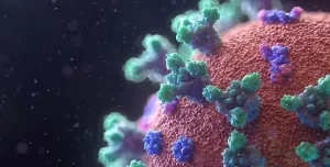
Here we’ve addressed common applications in infectious disease research including cell line development, binding affinity, viral neutralization, viral titer and more with a focused effort on understanding the SARS-CoV-2 virus in order to develop potential therapies for COVID-19 including vaccines, therapeutics and diagnostics.
Cardiomyocytes

Stem cells differentiated into cardiomyocytes are used to screen early for potential toxicological effects of drugs, thus helping to avoid investment in development of drugs which will fail in clinical trials due to cardiac toxicity.
Cell Counting

Cell counting is fundamental and critical to numerous biological experiments. Assays such as drug compound toxicity, cell proliferation, and inhibition of cell division rely on the assessment of the number or density of cells in a well.
Digital Confocal

The ability of cells to adapt to their environment is mediated by highly controlled signaling pathways. In many cases, the initiation of these pathways by extracellular signals, such as hormones or cytokines, leads to the translocation of proteins from the cytoplasm to the nucleus. Once in the nucleus, these proteins can regulate the expression of target genes. Assays requiring precise identification of sub-cellular structures or molecules within different cellular compartments can benefit from the ability of image deconvolution to improve identification of the specific fluorescence signal against potentially high background caused by out-of-focus light. The ImageXpress Pico system with Digital Confocal 2D on-the-fly deconvolution is capable of measuring the translocation of proteins from the cytoplasm to the nucleus, like NF-κB translocation upon treatment with TNF-α. The enhancement in image resolution and maintenance of quantitative information from the image deconvolution leads to greater statistical significance in the analysis of nuclear translocation.
Disease Modeling

Disease model systems range in complexity and scale from simple 2D cell cultures to complex model organisms. While model organisms offer in vivo context, they are often costly and may not represent human biology. On the other hand, while traditional 2D cell culture systems have been used for many years, they have limitations in representing the complex three-dimensional structure and cellular interactions found in living tissues. As a result, 3D cell cultures have emerged as an attractive model system for disease modeling.
Drug Discovery & Development

For every drug that makes it to the finish line, another nine don’t succeed. This alarming failure rate can be traced to reliance on 2D cell cultures that don’t closely mimic complex human biology, often leading to inaccurate predictions of a drug’s potential and extended drug development timelines.
Live cell imaging

Live cell imaging is the study of cellular structure and function in living cells via microscopy. It enables the visualization and quantitation of dynamic cellular processes in real time.
Live cell imaging encompasses a broad range of biological applications, from long-term kinetic assays to fluorescently labeling live cells.
Neurite Outgrowth / Neurite Tracing

Neurons create connections via extensions of their cellular body called processes. This biological phenomenon is referred to as neurite outgrowth. Understanding the signaling mechanisms driving neurite outgrowth provides valuable insight into neurotoxic responses, compound screening, and for interpreting factors influencing neural regeneration. Using the ImageXpress Micro system in combination with MetaXpress Image Analysis Software automated neurite outgrowth imaging and analysis is possible for slide or microplate-based cellular assays.
Oncology – Cancer Research

Cancer researchers need tools that enable them to more easily study the complex and often poorly understood interactions between cancerous cells and their environment, and to identify points of therapeutic intervention. Learn about instrumentation and software that facilitate cancer research using, in many cases, biologically relevant 3D cellular models like spheroids, organoids, and organ-on-a-chip systems that simulate the in vivo environment of a tumor or organ.
Stem Cell Research

Pluripotent stem cells can be used for studies in developmental biology or differentiated as a source for organ-specific cells and used for live or fixed cell-based assays on slides or in multi-well plates. The ImageXpress system has utility in all parts of the stem cell researcher’s workflow, from tracking differentiation, to quality control, to measuring functionality of specific cell types.
Toxicology

Toxicology is the study of adverse effects of natural or man-made chemicals on living organism. It is a growing concern in our world today as we are exposed to more and more chemicals, both in our environment and in the products we use.










 VI
VI