Fluorescence imaging platform within reach of every lab
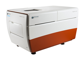
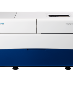
The ImageXpress® Nano Automated Imaging System features a long life, solid state, light engine, and optics to reliably deliver the right assay sensitivity. Capture fine details of a variety of cellular and subcellular assays with this powerful and flexible fluorescent microscopy solution. The system includes MetaXpress® High-Content Image Acquisition and Analysis Software with tools for 2D and 3D imaging and time lapse analysis, as well as a range of needs from ease-of-use through to proprietary assay design.
Image label-free
Brightfield imaging allows for rapid acquisition without the use of harmful fluorescent agents.
Streamline image analysis
The modular toolbox in the MetaXpress® software allows for the quick setup of hundreds of routine assays. Choose from our optional selection of turnkey application modules for greater convenience.
Capture a diverse range of samples
With 2x to 60x magnification, the system offers the flexibility to image whole-well (C. elegans, zebrafish), as well as sub-cellular details (vesicles, organelles).

MetaXpress high-content image aquisition and analysis software
- Meet high throughput requirements with a scalable, streamlined workflow
- Adapt your analysis tools to tackle your toughest problems, including 3D analysis
- Schedule automatic data transfer between third-party hardware sources and secure database
- Set up hundreds of routinely used HCS assays using MetaXpress software modules
Large field of view
An entire well of a 384-well plate can be captured in a single image at 4x magnification for faster throughput.
Automated Stages
Fully automated X, Y, and Z stages with resolution better than 25 nm.
Wide range of filters and objectives
The system can be configured with different filters or objectives (2-60x) to meet research needs.
Five fluorescent channels
The system can have up to 5 fluorescent filters installed at one time. The software allows up to 7 channels to be acquired at one time which enables multi-channel fluorescent and transmitted light imaging in one experiment.
High-speed autofocus
Laser autofocus enables quick, consistent focusing across plates, slides and uneven surfaces.
Environmental control option
Multi-day, time lapse and live cell assays can be run using the onboard environmental system with options for temprature, humidity, and CO2 control.
3D Cell Models

3D cell culture models offer the advantage of closely recapitulating aspects of human tissues including the architecture, cell organization, cell-cell and cell-matrix interactions, and more physiologically-relevant diffusion characteristics. Utilization of 3D cellular assays adds value to research and screening campaigns, spanning the translational gap between 2D cell cultures and whole-animal models. By reproducing important parameters of the in vivo environment, 3D models can provide unique insight into the behavior of stem cells and developing tissues in vitro.
COVID-19 and Infectious Disease Research
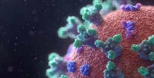
Here we’ve addressed common applications in infectious disease research including cell line development, binding affinity, viral neutralization, viral titer and more with a focused effort on understanding the SARS-CoV-2 virus in order to develop potential therapies for COVID-19 including vaccines, therapeutics and diagnostics.
Cardiomyocytes

Stem cells differentiated into cardiomyocytes are used to screen early for potential toxicological effects of drugs, thus helping to avoid investment in development of drugs which will fail in clinical trials due to cardiac toxicity.
Cell Counting

Cell counting is fundamental and critical to numerous biological experiments. Assays such as drug compound toxicity, cell proliferation, and inhibition of cell division rely on the assessment of the number or density of cells in a well.
Cell Migration Assays

The movement or migration of cells is often measured in vitro to elucidate the mechanisms of various physiological activities such as wound healing or cancer cell metastasis. Cell migration assays may be conducted in a controlled environment using live cell time lapse imaging. A “wound” in a confluent monolayer of cells growing in a microplate is created, either by manually creating a scratch or by utilizing special microplates that provide a uniform and reproducible cell-free zone. Monitor cell proliferation, wound healing, migration, and spreading using transmitted light or live cell-compatible fluorescence. These medium to high throughput assays may be used to compare the migration between cells treated with either inhibitory or stimulatory compounds.
Disease Modeling

Disease model systems range in complexity and scale from simple 2D cell cultures to complex model organisms. While model organisms offer in vivo context, they are often costly and may not represent human biology. On the other hand, while traditional 2D cell culture systems have been used for many years, they have limitations in representing the complex three-dimensional structure and cellular interactions found in living tissues. As a result, 3D cell cultures have emerged as an attractive model system for disease modeling.
Drug Discovery & Development

For every drug that makes it to the finish line, another nine don’t succeed. This alarming failure rate can be traced to reliance on 2D cell cultures that don’t closely mimic complex human biology, often leading to inaccurate predictions of a drug’s potential and extended drug development timelines.
Live cell imaging

Live cell imaging is the study of cellular structure and function in living cells via microscopy. It enables the visualization and quantitation of dynamic cellular processes in real time.
Live cell imaging encompasses a broad range of biological applications, from long-term kinetic assays to fluorescently labeling live cells.
Neurite Outgrowth / Neurite Tracing

Neurons create connections via extensions of their cellular body called processes. This biological phenomenon is referred to as neurite outgrowth. Understanding the signaling mechanisms driving neurite outgrowth provides valuable insight into neurotoxic responses, compound screening, and for interpreting factors influencing neural regeneration. Using the ImageXpress Micro system in combination with MetaXpress Image Analysis Software automated neurite outgrowth imaging and analysis is possible for slide or microplate-based cellular assays.
Oncology – Cancer Research

Cancer researchers need tools that enable them to more easily study the complex and often poorly understood interactions between cancerous cells and their environment, and to identify points of therapeutic intervention. Learn about instrumentation and software that facilitate cancer research using, in many cases, biologically relevant 3D cellular models like spheroids, organoids, and organ-on-a-chip systems that simulate the in vivo environment of a tumor or organ.
Stem Cell Research

Pluripotent stem cells can be used for studies in developmental biology or differentiated as a source for organ-specific cells and used for live or fixed cell-based assays on slides or in multi-well plates. The ImageXpress system has utility in all parts of the stem cell researcher’s workflow, from tracking differentiation, to quality control, to measuring functionality of specific cell types.
Toxicology

Toxicology is the study of adverse effects of natural or man-made chemicals on living organism. It is a growing concern in our world today as we are exposed to more and more chemicals, both in our environment and in the products we use.








 VI
VI