Capture large 3D organoid and spheroid images with up to double the speed
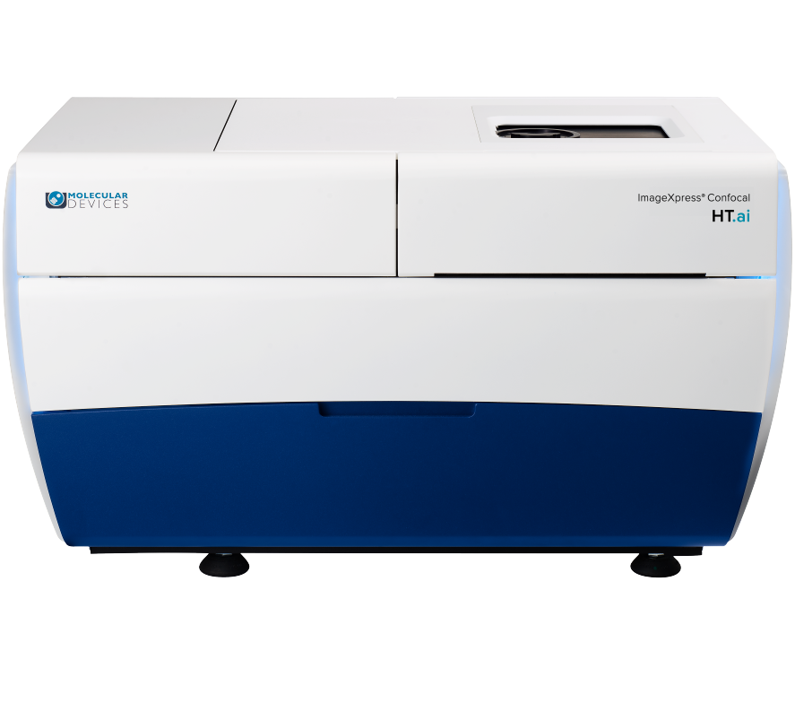
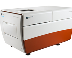
The ImageXpress® Confocal HT.ai High-Content Imaging System utilizes a seven-channel laser light source with eight imaging channels to enable highly multiplexed assays while maintaining high throughput by using shortened exposure times. Water immersion objectives improve image resolution and minimize aberrations so scientists can see deeper into thick samples.
The powerful combination of MetaXpress® software and IN Carta® software simplifies workflows for advanced phenotypic classification and 3D image analysis with machine learning capabilities and an intuitive user interface.
Enable greater assay flexibility
Eight imaging channels with laser excitation enable more assay flexibility, higher image brightness, and flexibility to use targeted imaging such as QuickID. Automated Water Immersion objectives offer greater numerical aperture and matched refractive index between the sample and the immersion media for enhanced resolution and decreased aberrations.
Increase throughput with higher quality images
Higher excitation power provides increased signal, shorter exposure times, and faster acquisitions of 3D samples. Micro-lens enhanced spinning disk confocal provides a flat field of view for more accurate and reproducible image analysis. Shorter exposure times generates up to a two-fold boost in scan speed. FRET experiments using lasers for CFP and YFP expand research.
Accelerate analysis speeds
IN Carta Image Analysis Software performs complex segmentation and classification. Phenoglyphs provides a robust trainable classification, and SINAP provides trainable segmentation for any image type. Accelerate analysis speeds by 40X with multi-threaded, parallel processing with MetaXpress® PowerCore Software. Reduces time from hours to minutes, eliminating 3D analysis as a bottleneck.

Images taken with same exposure times show different average intensities
IN Carta image analysis software

Powerful analytics combined with an intuitive user interface simplify workflows for image analysis and phenotypic profiling. Advanced features provide the functionality you need to analyze data in 2D, 3D, and 4D – at scale – and deliver real-time insights without the need for complex pre- or post- processing operations. Improve specificity of your image analysis workflows by utilizing the SINAP deep-learning module and see for yourself that Segmentation Is Not A Problem. Put machine learning to work for you and perform complex phenotypic analysis within a user-friendly Phenoglyphs module.
Cellular Image Gallery

Molecular Devices provides flexible options for the ImageXpress Confocal HT.ai system to meet your research needs and to easily capture images from different sample formats, including hanging drops and in round or flat bottom plates, for monitoring cell health kinetics under environmental control, and more. With over 30+ years of imaging expertise, we can help you select the right options to ensure the best images for your assay.
Standard hardware options
Water immersion objectives
20X, 40X, and 60X water immersion objectives improve the geometric accuracy during acquisition and reduce light refraction for brighter intensity at lower exposure times.
Transmitted light tower
Our transmitted light tower enables acquisition of high contrast images for unstained cells which can be easily viewed or separated from background.
Environmental Control
Environmental control maintains temperature and humidity levels while minimizing evaporation for multi-day, live cell, time-lapse imaging.
Customization options
Molecular Devices can successfully tailor the ImageXpress Confocal HT.ai system to include customized software and hardware including the features described below, as well as integration of other lab components such as incubators, liquid handlers, and robotics for a fully automated workcell. With over 30 years of experience in the life science industry, you can count on us to deliver quality products and provide worldwide support.
Deep tissue penetrating confocal disk module
The deep tissue penetrating, confocal disk module reduces crosstalk to improve out-of-focus light suppression and penetrate deeper into tissue.
Turnkey, high-throughput long term kinetics
Schedule and image multiple plates over long periods of time while keeping consistent temperature, O2(Hypoxia), CO2, and humidity conditions. Expand live cell walk-away capacity to 200+ plates.
Scale up robotic automation
Increase throughput, eliminate human errors, maintain sterility, and achieve consistent sample handling. Modular automation design—components can be added in modules and are upgradeable.
Deep Tissue Penetrating, Confocal Disk Module
Specialized deep tissue penetrating, confocal disk module, combined with a laser light source, improves light penetrance for deeper tissue penetration, resulting in sharper images with improved resolution when imaging thick tissue samples†.
- Improve suppression of out-of-focus light
- Reduce haze (pinhole crosstalk)
- Penetrate deeper into thick tissue samples for sharper images
3D Cell Imaging and Analysis

Three-dimensional (3D) cell models are physiologically relevant and more closely represent tissue microenvironments, cell-to-cell interactions, and biological processes that occur in vivo. Now you can generate more predictive data by incorporating technologies like the ImageXpress system with the integrated 3D Analysis Module in MetaXpress® software. This single interface will enable you to meet 3D acquisition and analysis challenges without compromise to throughput or data quality, giving you confidence in your discoveries.
COVID-19 and Infectious Disease Research
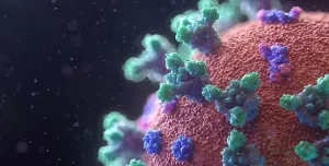
Here we’ve addressed common applications in infectious disease research including cell line development, binding affinity, viral neutralization, viral titer and more with a focused effort on understanding the SARS-CoV-2 virus in order to develop potential therapies for COVID-19 including vaccines, therapeutics and diagnostics.
Cell Painting

Cell Painting is a high-content, multiplexed image-based assay used for cytological profiling. In a Cell Painting assay, up to six fluorescent dyes are used to label different components of the cell including the nucleus, the endoplasmic reticulum, mitochondria, cytoskeleton, the Golgi apparatus, and RNA. The goal is to “paint” as much of the cell as possible to capture a representative image of the whole cell.

One common way of culturing cells in three dimensional space is through the use of extracellular matrix-based hydrogels, such as Matrigel. Cells are grown in an extracellular matrix (ECM) to mimic an in vivo environment. Differences between Matrigel and 2D cell cultures can be readily seen by their different cell morphologies, cell polarity, and/or gene expression. Hydrogels can also enable studies on cell migration and 3D structure formation, such as endothelial cell tube formation in angiogenesis studies.
Disease Modeling

Disease model systems range in complexity and scale from simple 2D cell cultures to complex model organisms. While model organisms offer in vivo context, they are often costly and may not represent human biology. On the other hand, while traditional 2D cell culture systems have been used for many years, they have limitations in representing the complex three-dimensional structure and cellular interactions found in living tissues. As a result, 3D cell cultures have emerged as an attractive model system for disease modeling.
Drug Discovery & Development

For every drug that makes it to the finish line, another nine don’t succeed. This alarming failure rate can be traced to reliance on 2D cell cultures that don’t closely mimic complex human biology, often leading to inaccurate predictions of a drug’s potential and extended drug development timelines.
Live cell imaging

Live cell imaging is the study of cellular structure and function in living cells via microscopy. It enables the visualization and quantitation of dynamic cellular processes in real time.
Live cell imaging encompasses a broad range of biological applications, from long-term kinetic assays to fluorescently labeling live cells.
Neurite Outgrowth / Neurite Tracing

Neurons create connections via extensions of their cellular body called processes. This biological phenomenon is referred to as neurite outgrowth. Understanding the signaling mechanisms driving neurite outgrowth provides valuable insight into neurotoxic responses, compound screening, and for interpreting factors influencing neural regeneration. Using the ImageXpress Micro system in combination with MetaXpress Image Analysis Software automated neurite outgrowth imaging and analysis is possible for slide or microplate-based cellular assays.
Oncology – Cancer Research

Cancer researchers need tools that enable them to more easily study the complex and often poorly understood interactions between cancerous cells and their environment, and to identify points of therapeutic intervention. Learn about instrumentation and software that facilitate cancer research using, in many cases, biologically relevant 3D cellular models like spheroids, organoids, and organ-on-a-chip systems that simulate the in vivo environment of a tumor or organ.
Organ-on-a-Chip

Organ-on-a-chip (OoC) is a technology that uses microfabrication techniques to create miniature models of biological organs, such as the lung, heart, or gut, on a chip-sized device. These microfabricated devices are made up of living cells that are grown on a microscale platform and mimic the structure and function of the organ they represent. The cells are typically arranged in a way that mimics the native three-dimensional structure of the organ and is perfused with fluids, such as blood or air, to represent the physiological environment of the organ.
Organoids

Organoids are three-dimensional (3D) multi-cellular microtissues that are designed to closely mimic the complex structure and functionality of human organs. Organoids typically consist of a co-culture of cells which demonstrate a high order of self-assembly to allow for an even better representation of complex in vivo cell responses and interactions, as compared to traditional 2D cell cultures.
Spheroids

Spheroids are multi-cellular 3D structures that mimic in vivo cell responses and interactions. They can be highly reproducible and to be scaled for high-content screening. Compared with adherent cells grown in 2D monolayers, 3D growth conditions are believed to more closely reflect the natural environment of cancer cells. Acquiring measurements from these larger structures involve acquiring images from different depths (z-planes) within the body of the spheroid and analyzing them in 3D, or collapsing the images into a single 2D stack before analysis.
Stem Cell Research

Pluripotent stem cells can be used for studies in developmental biology or differentiated as a source for organ-specific cells and used for live or fixed cell-based assays on slides or in multi-well plates. The ImageXpress system has utility in all parts of the stem cell researcher’s workflow, from tracking differentiation, to quality control, to measuring functionality of specific cell types.
Toxicology

Toxicology is the study of adverse effects of natural or man-made chemicals on living organism. It is a growing concern in our world today as we are exposed to more and more chemicals, both in our environment and in the products we use.









 VI
VI