The SpectraMax® M Series Multi-Mode Microplate Readers measure UV and visible absorbance, fluorescence, luminescence, fluorescence polarization, TRF and HTRF. Standard features include a cuvette port, spectral scanning in 1 nm increments, and up to six wavelengths per read. These robust readers have been placed in labs from Antarctica to the International Space Station. With optimized reagents, validation tools, and industry-leading SoftMax® Pro Software, they provide consistent performance.
 Assay flexibility
The optical system uses two scanning monochromators so you can determine optimal excitation and emission settings, resulting in assay performance similar to that of dedicated single-mode readers.
 Better absorbance accuracy
Measure sample depth with no temperature dependency using the PathCheck Sensor technology. It automatically normalizes absorbance readings to 1 cm, eliminating the need for standard curves.
 Increased throughput
Integrate easily with our StakMax® Microplate Stacker for walk-away automation. The stacker can hold up to 50 plates and can be configured to include a barcode reader.
Kickstart your compliance journey with our “GxP Starter Kit”
For researchers working in GLP or GMP laboratories, achieve full GxP compliance with our turnkey solution:
- SpectraMax M-Series Microplate Reader are modular and upgradeable with a wide range of high performance capabilities.
- SoftMax® Pro 7.2 GxP Software – The latest, most secure software to achieve full FDA 21 CFR Part 11 and EudraLex Annex 11 compliance with streamlined workflows to ensure data integrity.
- SoftMax® Pro Validation Package – Provides the most comprehensive documentation and tools available to validate GxP administrator features, software operation, and analysis functions for microplate reader instrumentation.
- Software Installation & Training – Our software installation services verify and document that required components are installed to operational specifications.
Specifications & Options of SpectraMax M Series Multi-Mode Microplate Readers
|
M2/M2e Multi-Mode Microplate Reader
|
M3 Multi-Mode Microplate Reader
|
M4 Multi-Mode Microplate Reader
|
M5/M5e Multi-Mode Microplate Reader
|
| General specifications |
| Read modes |
Absorbance
Fluorescence (top/bottom read) |
Absorbance
Fluorescence (top/bottom read)
Luminescence (top/bottom read) |
Absorbance
Fluorescence (top/bottom read)
Luminescence (top/bottom read)
Time-Resolved Fluorescence (top/bottom read) |
Absorbance
Fluorescence (top/bottom read)
Luminescence (top/bottom read)
Time-Resolved Fluorescence (top/bottom read)
Fluorescence Polarization (top read)
HTRF (top/bottom read) |
| Wavelength ranges |
Abs: 200 – 1000 nm FL Ex: 250 – 850 nm FL Em: 360 – 850 nm (M2) FL Em: 250 – 850 nm (M2e) |
Abs: 200 – 1000 nm FL: 250 – 850 nm Lumi: 250 – 850 nm |
Abs: 200 – 1000 nm FL: 250 – 850 nm Lumi: 250 – 850 nm TRF: 250 – 850 nm |
Abs: 200 – 1000 nm FL: 250 – 850 nm Lumi: 250 – 850 nm TRF: 250 – 850 nm FP: 300 – 750 nm |
| Wavelength selection |
Monochromator tunable in 1-nm increments |
Monochromator tunable in 1-nm increments |
Monochromator tunable in 1-nm increments |
Monochromator tunable in 1-nm increments |
| Absorbance photometric accuracy/linearity |
< ±1.0% and ±0.006 OD (0.0-2.0 OD) |
< ±1.0% and ±0.006 OD (0.0-2.0 OD) |
< ±1.0% and ±0.006 OD (0.0-2.0 OD) |
< ±1.0% and ±0.006 OD (0.0-2.0 OD) |
| Absorbance photometric precision/repeatability |
< ±1.0% and ±0.003 OD (0.0-2.0 OD) |
< ±1.0% and ±0.003 OD (0.0-2.0 OD) |
< ±1.0% and ±0.003 OD (0.0-2.0 OD) |
< ±1.0% and ±0.003 OD (0.0-2.0 OD) |
| Sensitivity top read optimized |
FL: 3.0 fmol/well in 200 μL FITC 96 well (15 pM), 3.0 fmol/well in 75 μL FITC 384 well (40 pM) |
FL: 1 fmol/well in 200 μL FITC 96 well (< 5 pM), 2 fmol/well in 100 μL FITC 384 well (< 20 pM) Lumi: < 2 fg/well for firefly luciferase in 96 and 384 well |
FL: 1 fmol/well in 200 μL FITC 96 well (< 5 pM), 2 fmol/well in 100 μL FITC 384 well (< 20 pM) Lumi: < 2 fg/well for firefly luciferase in 96- and 384-well TRF: 100 fM europium in 96 or 384 well |
FL: 1 fmol/well in 200 μL FITC 96 well (< 5 pM), 2 fmol/well in 100 μL FITC 384 well (< 20 pM) Lumi: < 2 fg/well for firefly luciferase in 96 and 384 well TRF: 100 fM europium in 96 and 384 well FP: < 5 mP standard deviation at 1 nM fluorescein in 96 and 384 well |
| Light source(s) |
Xenon flashlamp |
Xenon flashlamp |
Xenon flashlamp |
Xenon flashlamp |
| Detector(s) |
Silicon photodiode
Photomutiplier tube |
Silicon photodiode
Photomutiplier tube |
Silicon photodiode
Photomutiplier tube |
Silicon photodiode
Photomutiplier tube |
| Read types (endpoint, kinetic, etc) |
Endpoint
Kinetic
Spectrum scan
Well scan |
Endpoint
Kinetic
Spectrum scan
Well scan |
Endpoint
Kinetic
Spectrum scan
Well scan |
Endpoint
Kinetic
Spectrum scan
Well scan |
| Read times |
Abs: 17 seconds
FL: 15 seconds |
Abs: 17 seconds
FL: 15 seconds
Lumi: 17 seconds |
Abs: 17 seconds
FL: 15 seconds
Lumi: 17 seconds
TRF: 15 seconds |
Abs: 17 seconds
FL: 15 seconds
Lumi: 17 seconds
TRF: 15 seconds
FP: 44 seconds |
| Plate shaking |
Linear |
Linear |
Linear |
Linear |
| Cuvette port |
 |
 |
 |
 |
| Robotics / automation |
 |
 |
 |
 |
| Plate type(s) |
6 – 384 well plates |
6 – 384 well plates |
6 – 384 well plates |
6 – 384 well plates |
| Temperature control |
 Ambient + 4°C to 45°C Ambient + 4°C to 45°C |
 Ambient + 2°C to 60°C Ambient + 2°C to 60°C |
 Ambient + 2°C to 60°C Ambient + 2°C to 60°C |
 Ambient + 2°C to 60°C Ambient + 2°C to 60°C |
| Dimensions |
H 22 cm x W 58 cm x D 39 cm |
H 22 cm x W 58 cm x D 39 cm |
H 22 cm x W 58 cm x D 39 cm |
H 22 cm x W 58 cm x D 39 cm |
| Validation tools |
 Absorbance validation plate Absorbance validation plate
Fluorescence validation plate
SoftMax Pro GxP Software |
 Absorbance validation plate Absorbance validation plate
Fluorescence validation plate
Luminescence validation plate
SoftMax Pro GxP Software |
 Absorbance validation plate Absorbance validation plate
Fluorescence validation plate
Luminescence validation plate
SoftMax Pro GxP Software |
 Absorbance validation plate Absorbance validation plate
Fluorescence validation plate
Luminescence validation plate
SoftMax Pro GxP Software |
Absorbance

Cell Health

Cell viability refers to the number of healthy cells in a population and can be evaluated using assays that measure enzyme activity, cell membrane integrity, ATP production, and other indicators. These methods can employ luminescent, fluorescent, or colorimetric readouts as indicators of general cell viability or even specific cellular pathways. Cytotoxicity and cell viability assays are often used to assess a drug or other treatment’s effect, and are valuable tools in the search for new therapeutics, as well as advancing our understanding of how normal cells function.
Cell viability

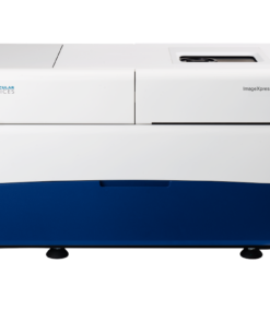
Cell line development requires the discovery of single cell-derived clones that produce high and consistent levels of the target therapeutic protein. A critical first step in the process is the isolation of single, viable cells. Single cells proliferate to form colonies that can then be assessed for productivity of the target therapeutic protein. Viability and growth rates of single cell-derived clones can be characterized on the CloneSelect Imager and the SpectraMax i3x microplate reader and imaging cytometer.
Cellular Signaling

Cellular signaling allows cells to respond to their environment as well as to communicate with other cells. Proteins located on the cell surface can receive signals from the surroundings and transmit information into the cell via a series of protein interactions and biochemical reactions that comprise a signaling pathway. Multicellular organisms rely upon an extensive array of signaling pathways to coordinate the proper growth, regulation, and functioning of cells and tissues. If signaling between or within cells is dysregulated, inappropriate cellular responses may lead to cancer and other diseases.
Drug Discovery & Development

For every drug that makes it to the finish line, another nine don’t succeed. This alarming failure rate can be traced to reliance on 2D cell cultures that don’t closely mimic complex human biology, often leading to inaccurate predictions of a drug’s potential and extended drug development timelines.
ELISA

Enzyme-linked immunosorbent assays (ELISAs) are used to measure the amount of a specific protein, using a microplate format, and results are most often detected via absorbance in the visible wavelength range. Chemiluminescent and fluorescent ELISA formats offer enhanced sensitivity for accurate quantitation of less abundant analytes.
Fluorescence Polarization (FP)
IgG Quantification

In therapeutic protein engineering and cell line development, IgG quantification is the process of measuring the amount of immunoglobulin G (IgG), a common class of therapeutic proteins, produced by a genetically modified cell line. This is important for evaluating and monitoring the productivity of the cell line and selecting the best candidate for further development of therapeutic antibodies.
Luminescence

Microbiology and Contaminant

Microbes, including bacteria, have been estimated to make up about 15 percent of the earth’s biomass, and microbes in the human body outnumber human cells by 10 to 1. These microorganisms provide great benefit to us and are also vital to many fields of research from medicine to alternative energy production. On the other hand, monitoring for microbes and the toxic substances they produce is necessary to ensure the safety of pharmaceutical products. Scientists whose research relies on mammalian cells must carefully monitor these cultures for unwanted microbial contaminants to ensure that their experimental results are reliable.
Nucleic Acid (DNA/RNA) Quantitation and Analysis

Nucleic acids are large biomolecules common to all known life forms. Deoxyribonucleic acid (DNA) consists of a double strand of pairs of nucleotides, while ribonucleic acid (RNA) is typically a single strand. In DNA, the nucleotides are adenine, cytosine, guanine, and thymine, while RNA contains uracil instead of thymine. DNA makes up the genetic material of all organisms, encoding the information cells need to synthesize proteins.
Protein Detection, Quantitation and Analysis

Protein detection, quantitation, and analysis are central to the investigation of a wide variety of biological processes. Measuring the concentration of protein is necessary to processes ranging from protein purification and labeling to sample preparation for electrophoresis. Protein can be quantitated directly via absorbance at 280 nm, or indirectly using colorimetric (BCA, Bradford, etc.) or fluorometric methods offering advantages such as greater sensitivity. To identify and measure a specific protein within a complex sample, for example, serum or cell lysate, an ELISA may be used.
SNP genotyping

Genotyping is a process for analyzing genetic differences among individuals by examining their DNA sequences. Single nucleotide polymorphisms (SNPs) are one of the most common types of genetic variation, consisting of a single nucleotide mutation at a specific locus. SNP genotyping has proven very useful in identifying disease-related mutations in various species, and as a result many techniques for SNP detection have been developed.
TRF, TR-FRET và HTRF

 Assay flexibility
 Better absorbance accuracy
 Increased throughput







 P
P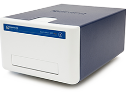


 VI
VI